At Altig Optical, we believe in providing the highest standard of care by using the latest advancements in eye care technology. Our commitment to cutting-edge tools and techniques ensures precise diagnostics, effective treatments, and an exceptional experience for every patient.
Whether you’re visiting for an eye exam in Fort Worth or exploring advanced solutions for dry eye or myopia management, our technology supports your vision and eye health.
Our State-of-the-Art Eye Care Technology
We are proud to offer a range of advanced diagnostic and treatment technologies to meet your unique vision needs:
Corneal Topography
Corneal topography, also known as photokeratoscopy or videokeratography, is a non-invasive medical imaging technique for mapping the surface curvature of the cornea, the outer structure of the eye. Since the cornea is normally responsible for some 70% of the eye's refractive power, its topography is of critical importance in determining the quality of vision.
The three-dimensional map is, therefore, a valuable aid to Dr. William D. Altig in the diagnosis and treatment of a number of conditions such as keratoconus and other corneal diseases. It is also very important for the fitting of special contact lenses and orthokeratology or VST (Vision Shaping Treatment). Corneal topography can also assist in planning refractive surgery such as LASIK and evaluation of its results.
Digital Retinal Imaging & OCT Scans at Altig Optical
This is very important in assisting Dr. Altig to detect and measure any changes to your retina each time you get your eyes examined, as many eye conditions, such as glaucoma, diabetic retinopathy, and macular degeneration are diagnosed by detecting changes over time.
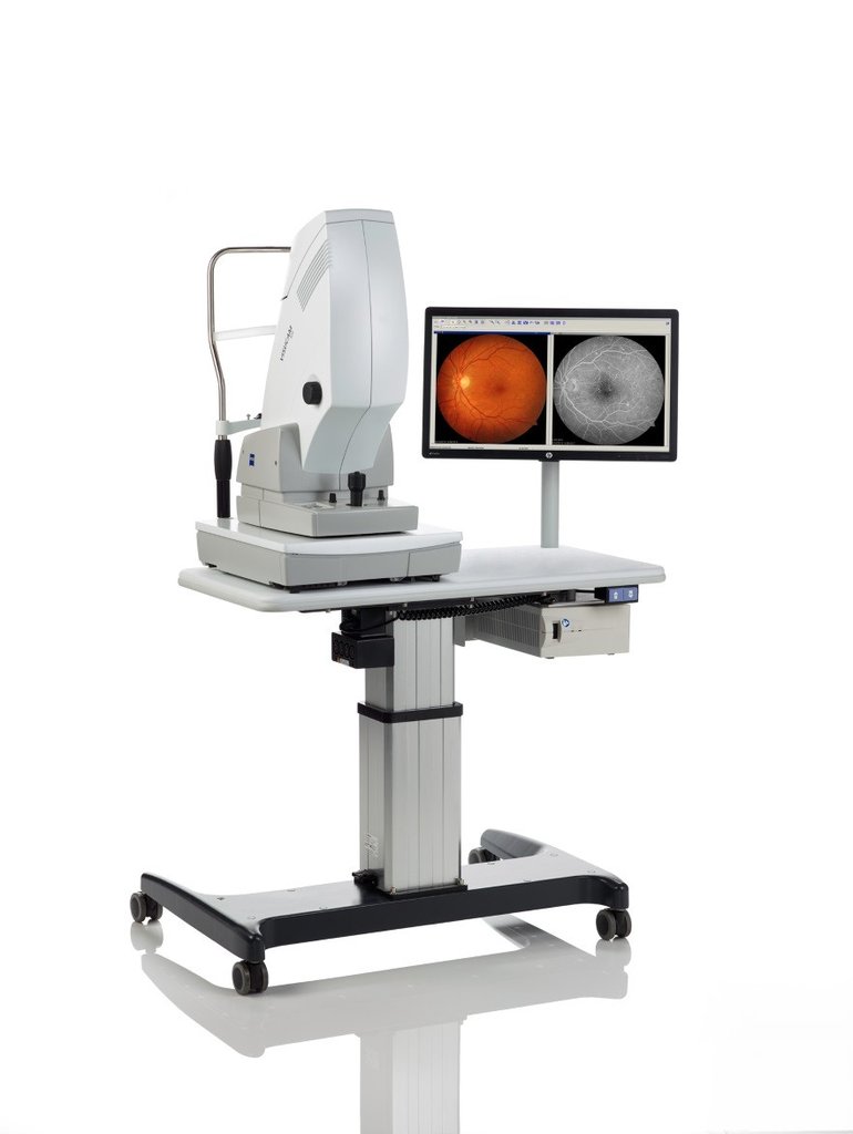
VISUCAM
Superior clarity, ultra-high resolution, revolutionary ZEISS optics.

Zeiss Visucam
We are proud to introduce the latest cutting-edge technology in retinal imaging, the ZEISS VISUCAM
VISUCAM retinal scan technology is now available at Altig Optical. This amazing device allows your eye doctor to see retinal, optic nerve and cornea structures not visible through regular exam methods by using light to provide a high-resolution scan meant to pick up early signs of structural change or disease.
Digital Retinal Imaging
Digital Retinal Imaging allows Dr. Altig to evaluate the health of the back of your eye, the retina. It is critical to confirm the health of the retina, optic nerve, and other retinal structures. The digital camera snaps a high-resolution digital picture of your retina. This picture clearly shows the health of your eyes and is used as a baseline to track any changes in your eyes in future eye examinations.
The advantages of digital imaging include:
- Quick, safe, non-invasive and painless
- Provides detailed images of your retina and sub-surface of your eyes
- Provides instant, direct imaging of the form and structure of eye tissue
- Image resolution is extremely high quality
- Uses eye-safe near-infra-red light
- No patient prep required
Optical Coherence Tomography (OCT)
An Optical Coherence Tomography scan (commonly referred to as an OCT scan) is the latest advancement in imaging technology. Similar to ultrasound, this diagnostic technique employs light rather than sound waves to achieve higher-resolution pictures of the structural layers of the back of the eye.
OCT provides detailed, cross-sectional images of your retina, allowing us to detect early signs of eye diseases like macular degeneration, diabetic retinopathy, and glaucoma. This non-invasive technology ensures accurate diagnoses and proactive care.
These detailed images are revolutionizing the early detection and treatment of eye conditions such as wet and dry age-related macular degeneration, glaucoma, retinal detachment, and diabetic retinopathy.
An OCT scan is a noninvasive, painless test. It is performed in about 10 minutes right in our office. Feel free to contact our office to inquire about an OCT at your next appointment.
Visual Field Testing With our Eye Doctor
A visual field test measures how much 'side' vision you have. It is a straightforward test, painless, and does not involve eye drops. Essentially lights are flashed on, and you have to press a button whenever you see the light. Your head is kept still and you have to place your chin on a chin rest. The lights are bright or dim at different stages of the test. Some of the flashes are purely to check you are concentrating.
Each eye is tested separately and the entire test takes 15-45 minutes.
The Visual Field Test is carried out by a computerized machine, called a Humphrey Visual Field Analyzer. For each test you have to look at a central point and then press a buzzer each time you see the light.
We now have a Matrix Visual Field Analyzer. This allows us to more quickly screen and test for glaucoma and other neurological illnesses.
Anterior Segment Camera
The anterior segment camera is an optometric device that is used to examine the front surface and anterior portion of the eye helps to detect and track the progression of external diseases of the eyelids, blepharitis, styes, corneal diseases, conjunctival diseases, and cataracts.
 It is a very important tool for documenting diseases of the eye as a baseline to watch for progression or changes associated with the conjuctiva, chronic lid conditions or other conditions of the conjuctiva and cornea such as pterygiums. It is also helpful when fitting specialty contact lenses, scleral lenses, gas permeable and hybrid lenses for the treatment of various corneal diseases such as keratoconus.
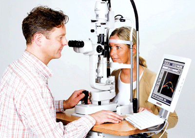
Describing the convenience and opportunities for patient education afforded by the anterior segment camera, Dr. Altig comments, “Since I'm already looking at the patient's eye with the slit lamp, it is very easy to take a picture at the same time, making it easy and comfortable.
The photo comes up on a large monitor when the person is still sitting in the exam chair so it is easy to explain to the patient what I am looking at. This is also true with contact lenses. With specialty contact lenses, it is easy to explain the fit and reason for larger scleral lenses.”
Dr. Altig recommends that the anterior segment camera be used for patients who need to see what is going on with conditions such as blepharitis, when lid hygiene is important, to demonstrate to the patient how lid hygiene and lid scrubs will help the condition. This is also true with conditions such as nevi and pterygiums, which often need to be monitored for progress, making a baseline photo a valuable tool.
For more information about the anterior segment camera, contact Dr. Altig today.
Fundus Photography
Precise Diagnosis of Diabetes & Eye Disease
 Fundus photography uses a specialized, sophisticated digital camera with powerful lenses that focus on the ocular structures of your inner eye. Your fundus is found at the back of your eyeball, which is where your retina is located. A classic fundus photo will show your optic nerve, main retinal blood vessels, and macula. These images are used to record and diagnose many eye conditions, including inspecting closely for the effects of diabetes.
At Altig Optical our office is equipped with Canon’s Retinal Camera with advanced fundus photography. Dr. Altig, our professional optometrist, is skilled and experienced in recording and analyzing these pictures as a part of our comprehensive eye exams. We will document the images of your fundus and compare them during follow-up visits to our Fort Worth, TX, office. This is a highly effective way to detect changes in your eye health, especially diabetic eye conditions, as well as pay attention to the retinal disease, macular holes, macular degeneration, and retinal detachment or tears.
How does fundus photography work?
Dilating eye drops will first be placed in your eyes, as light rays from the fundus camera enter your eye through the pupil. After the drops take effect, we will ask you to sit at our compact Canon camera, resting on the chin rest with your forehead against the bar. Once we align and focus the camera, you’ll see a quick flash of light, which generates the fundus photograph.
What eye conditions can be detected with fundus photography?
A fundus photo shows our Scarsdale eye doctors a comprehensive, detailed view of your retina. By comparing images from a visit to visit, any changes in your retinal health will be noticed efficiently. In particular, Dr. Altig uses fundus photography to keep watch on the signs of diabetes.
Recorded fundus pictures are ideal for tracking the progression of diabetic retinopathy, in which uncontrolled diabetes damages the retina, or macular swelling (macular edema). We will also pay attention to the effects of glaucoma on the optic nerve. In comparison to direct eye examinations, fundus photography makes many of the ocular symptoms caused by diabetes easier to visualize.
We’ll use the latest diagnostics, such as fundus photography, to evaluate your diabetes and ocular health during a comprehensive eye examination at Altig Optical. We’re proud to serve patients from Fort Worth, Newark, North Richland Hills, Saginaw, and Watauga, with state-of-the-art eye care! Dr. Altig looks forward to seeing you – call to schedule your appointment.
